Farrier Takeaways

  • White line disease is an opportunistic, microbial infection that leads to degradation of the non-pigmented stratum medium layer of the hoof. It is a common disorder in many regions, and although most cases are relatively minor, severe cases can develop.
  • Severe cases involving coffin bone rotation should be differentiated from other causes of displacement, such as laminitis.
  • Cases with separation and cavities of the hoof wall usually require hoof wall resection to facilitate proper treatment.

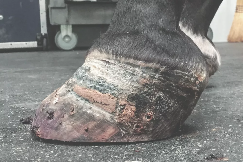
White line disease (WLD) is a common hoof disorder, particularly in horses kept in moist or humid environments. While many cases are relatively minor, severe cases can develop that require the expertise of a farrier and veterinarian team.

WLD is a microbial infection of the non-pigmented stratum medium layer of the hoof wall. The infection is a result of a mixed-population of opportunistic bacteria and fungi that are keratinopathogenic and cause the degradation of keratin — a protein that composes the insensitive structures of the hoof. This degradation creates separation and cavities within the layers of the hoof that can span from a few millimeters deep to the entire height of the hoof wall. These microbes are believed to be present in the environment and infect the non-pigmented stratum medium when provided with the appropriate conditions.

Although extremely common in some regions, the majority of cases are relatively mild and can be managed with routine trimming, improved hygiene and topical medications. However, severe cases can develop that result in lameness and extensive damage to the hoof, and require aggressive treatment.

The patient is an 11-year-old American Quarter Horse Association mare that is used for Western Pleasure showing and riding. She has a documented history of “high-low” forefeet conformation, with the left being more upright. She was purchased by her current owners 2 years ago with no prior history of forelimb lameness and has lived in the humid continental climate of central Indiana. She had been maintained on a consistent 6-week shoeing cycle and was in regular work.

Left forefoot at presentation.


You May Also Be Interested In...

White Line Disease: Different Approaches To An Old Problem

Now is the time to learn how to protect your clients’ horses against one of the more troublesome aspects of quality hoof care. Our in-depth eGuide on white line disease aims to dramatically help the horses in your practice suffering from this ailment, and it’s FREE! Download now »


She reportedly developed a left forelimb lameness while at a horse show 2 weeks prior. She presented to Janssen Veterinary Clinic, in Sheridan, Ind., for a lameness evaluation when there was no improvement in her lameness following being reshod.

The horse reportedly exhibited a consistent left forelimb lameness at the trot. At presentation, she was sound at the walk. She was not trotted due to concerns over the stability of the left front hoof capsule. The left forefoot had a more upright conformation than the right. The dorsal hoof wall had a dished appearance and the wall sounded hollow when percussed. The sole was convex, giving it a “dropped” appearance (Figures 1a and 1b) and she was reactive to hoof testers across the toe. She was shod with open-heeled steel shoes on both front feet. When the left shoe was removed, there was significant separation of the hoof wall at the toe and quarters.

Radiographs of the left forefoot re­vealed a large gas opacity undermining the dorsal hoof wall, consistent with severe WLD resulting in hoof capsule destabilization and coffin bone rotation (Figure 2). Sole depth was decreased at 8mm. The right forefoot was comparatively normal (Figure 3).

Radiograph of left forefoot at presentation.

Radiograph of right forefoot at presentation.

Hoof Capsule Destabilization And Coffin Bone Displacement

While coffin bone rotation can be a sequela of severe WLD, it’s important to differentiate it from other causes of displacement, namely laminitis. Although the resulting rotation may look similar on radiographs, the failure within the hoof capsule occurs at different locations.

As previously mentioned, WLD affects the non-pigmented stratum medium. Thus, hoof capsule separation and eventual destabilization occur at the stratum medium-stratum internum junction. Laminitis affects the lamellae themselves, and separation occurs at the dermal and epidermal lamellar interface.

Since prognosis and treatment for laminitis and WLD are vastly different, an accurate diagnosis for the cause of the coffin bone displacement is paramount. It is worth mentioning that horses with laminitis can develop WLD, but they are not always comorbidities.

While the exact etiology of WLD has not been proven, there are a host of contributing factors that seem to predispose a horse to develop WLD.

The left forefoot of this patient re­portedly has always been more up­right. This conformation places ex­cess strain on the dorsal aspect of the hoof wall, leading to overloading and stretching of the white line.

The climate of central Indiana is humid with ample annual rainfall. This leads to wet, muddy conditions, which soften hooves and seem to favor growth of the causative fungi and bacteria.

The patient has no known history of laminitis. She had a healthy body condition at presentation, and there were no real concerns with her other three feet, thus laminitis was not determined to be a contributing factor to this case of WLD.

Debris within the hoof wall separation found at time of resection.

Initial Treatment/Hoof Wall Resection

The mainstay of treating WLD is to expose the causative microorganisms to ultraviolet light, air and topical antiseptics. It is particularly important to treat the proximal aspect of affected areas, as this is where the disease propagates. In horses with large defects, treating from only the solar aspect is rarely rewarding as the proximal margin is often insufficiently affected. In these cases, hoof wall resection is warranted to allow proper exposure of the tissues.

That was the course of treatment pursued for this patient. All undermined hoof wall was resected. Within the defect, there were large accumulations of white and black, crumbly, keratin debris that is characteristic of the disease (Figure 4). Edges of the defect were debrided using a Dremel until healthy appearing margins were reached. Since WLD is restricted to avascular, insensitive tissues, there was no hemorrhage or pain associated with the procedure. Radiographs following the hoof wall resection highlight the degree of tissue that needed to be debrided (Figures 5a and 5b).

Left forefoot following hoof wall resection.

The topical an­ti­septic utilized in this case was a 7% strong tincture of io­dine. In addition to killing bacteria and fungus, the alcohol base helps keratinize and harden the areas of hoof it is applied to. Hardening these tissues acts as a mechanical barrier, making it more difficult for microbes to penetrate and reinfect these tissues.

At the time of resection, the patient’s left forefoot was trimmed with the goal of reducing palmar angle while retaining all sole depth. The patient was comfortable at the walk before and after the hoof wall resection, and care was taken to not induce soreness by trimming away too much heel height at one time. A more normal coffin bone alignment was achieved by incrementally trimming away heel height over the course of several weeks, which allowed her to gradually acclimate to the change and remain clinically comfortable.

LEARN MORE

Here are more ways to gain insight about white line disease, including::

  • Watching the Online Hoof-Care Classroom, “Best Tips For
    Managing Springtime Footcare.”
  • Reading, “How Nutrition
    Impacts White Line Disease.”
  • Downloading the free eGuide, “White Line Disease: Different Approaches To An Old
    Problem.”
Access this content by visiting
americanfarriers.com/0418

Initially, joint appointments with the farrier and veterinarian team met at 3-week intervals. During these appointments, radiographs of the left forefoot were obtained and used to guide trimming decisions. The hoof wall defect was lightly debrided with a Dremel to ensure all margins were still healthy appearing and there were no areas of recurring WLD. Once she had returned to a more normal coffin bone alignment and the team was confident that the WLD was under control, she was maintained on a 6-week trimming cycle. Radiographs were repeated every 12 to 18 weeks to ensure recovery was progressing as desired.

It was agreed upon that this horse would require some form of solar protection from the ground. Due to the amount of hoof wall resected away, nail-on shoes were not feasible. Hoof boots or glue on shoes were her best options. Therapeutic (SoftRide brand) hoof boots were chosen due to farrier comfort, and the ability to remove the boot and visualize and treat the entire hoof as needed. The gel orthotic provided a soft surface for her to stand on, as well as load sharing with the frog and sole to reduce force placed on the remaining hoof wall. The boots were worn at all times during the first few weeks of treatment. As the case progressed, she was allowed to go barefooted while in a bedded, clean stall.

As previously discussed, WLD seems more likely to occur in dirty or wet conditions. Thus, husbandry changes were enacted in an effort to keep her feet as clean and dry as possible while the hoof wall defect grew out. The hoof boots themselves do an excellent job protecting the hooves and keeping them away from ground contaminants. The owner was instructed to keep the patient’s stall as clean as possible, especially while stalled barefoot. Turnout was to be in dry (not muddy) conditions only. Initially, the hoof wall defect was cleaned with a wire brush and treated daily with the 7% strong tincture of iodine. Once the WLD was under control, treatment was decreased to two to three times weekly.

Once the left coffin bone alignment and WLD had improved, the option of glue-on shoes was revisited so that the patient could return to light work. The owner was concerned with making changes since treatment had thus far been successful, so it was decided to continue keeping the patient barefoot with therapeutic hoof boots until the defect fully grew out.

Almost 10 months after the hoof wall resection, the last of the hoof wall defect was trimmed away. The patient was sound, and her left forefoot conformation had returned to her baseline normal (Figure 6).

Left forefoot at resolution of the case.

Future Management

Four months following resolution of the case, the patient remained sound and her hoof capsule remained healthy. However, given her history and predisposing factors, measures should always be taken to prevent recurrence.

The solar aspect of the hoof should be protected from mechanical trauma, which could lead to weakened areas for microbes to invade. The patient historically has always been shod while working. If the owner elects to keep the patient barefooted, use of hoof boots during turnout is encouraged and exposure to muddy conditions should be limited as much as possible.

Ideally, an antimicrobial environment would be maintained between the shoe and hoof. At the moment, a commercial copper sulfate paste is applied along the white line prior to the shoe being nailed on. An alternative would be to apply a full pad with antiseptic, aerobic packing beneath.

While regular joint appointments for the patient’s WLD are no longer needed, there remains an open line of communication between the farrier and veterinarian about her to ensure there are no concerns going forward.