
American Farriers Journal
American Farriers Journal is the “hands-on” magazine for professional farriers, equine veterinarians and horse care product and service buyers.
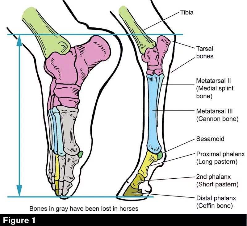
Homology study: the foot in humans vs. horses. These images are not to the same scale; rather, the feet have been reduced to equal length as measured from the point of the hock to the tip of the coffin bone. The human foot has been rotated about 90 degrees to put it in the stance normal to equines. Horsemen refer only to the structures surrounding the coffin bone as the horse’s “foot,” but the anatomical foot consists of everything between the horizontal blue lines. Color coding shows bones that are homologous (have the same embryological and evolutionary origin). An equivalent diagram for the forelimb was presented previously (AFJ, November, 2019 pp. 66-74).
In this series, Dr. Deb Bennett examines the equine hind limb. In this installment, Bennett explores the hock.
Weight-bearing upon flexed hocks puts enormous strain on all parts of the hock joint. Along with the equally complex stifle joint, the hock is crucial to the horse’s ability to flex and extend the hind limb and create the forward thrust that is the “impulsion” so often sought by horse owners who compete in the Olympic disciplines.
In the Western disciplines, the hock must stand up to sliding stops, rollbacks and bursting acceleration. Horses used for packing or trail riding must have hocks that will go all day, up and down steep hills, without rupture or fatigue.
The hock is structured by bones of peculiar shape. The tibial tarsal bone or astragalus upon whose oblique trochlea the distal end of the tibia rides also has a large shelf to support the deep digital flexor (DDF) tendon. The large fibular tarsal bone or calcaneus interlocks with the astragalus…
American Farriers Journal is the “hands-on” magazine for professional farriers, equine veterinarians and horse care product and service buyers.

American Farriers Journal is the “hands-on” magazine for professional farriers, equine veterinarians and horse care product and service buyers.
Download these helpful knowledge building tools


