Keratomas can appear cylindrical, looking like columns deep behind the hoof wall and oriented parallel to the horn tubules. These masses typically present as aberrant columnar-shaped proliferation and thickening of keratin within the horn that extends toward the inside of the hoof.1
In addition, spherical keratomas occur, even if they are less common and have been reported in the frog, in the sole and above the coronary band. There is still a lack of knowledge about the etiology of keratomas in horses. However, they seem to develop because of localized chronic irritation, inflammation or trauma to the germinal layers of the epithelium of the hoof.1,2,3 Therefore, keratomas often are seen in hoof regions with recurrent abscesses migrating through the lamellar layer of the inner hoof capsule or as the result of chronic, deep, or even bleeding hoof cracks.
The dermis and stratum germinativum of the epidermis's chronic irritation seems to cause a kind of malignant degeneration of the keratinocytes, comparable with tumor-like scar tissue. The tumor develops into an expansile mass that usually remains deep in the hoof capsule.
Keratomas are described histologically as rings of squamous epithelial cells containing abundant keratin.1,2 This poorly structured soft horn may be more susceptible to microbial colonization than is normal horn. Therefore, the keratoma can become chronically infected and develop into a purulent lesion.
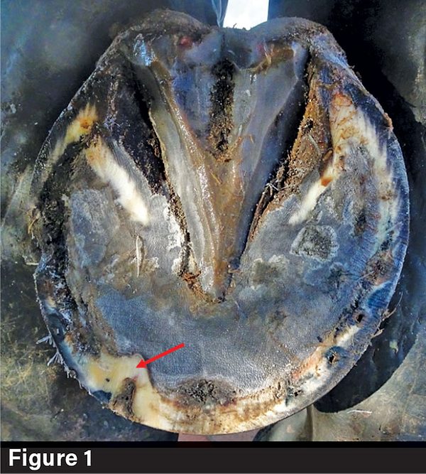
Often, the unstructured mass increases in size. This may exert pressure and cause lysis of the underlying distal phalanx or distortion of the overlying hoof capsule. A change in the white line’s contour is often the only initial clinical sign of a keratoma. It’s most often noticed by the farrier while trimming the foot, at a spot where the lamellar horn of the white line is replaced by tubular horn and scar tissue. This is visible on solar examination as a local, semicircular and thickened inward-bound white line (Figure 1).
Radiographic evidence of a keratoma can be a well-demarcated circular or oval area of lysis in the solar margin or parietal surface of the coffin bone. However, mild bone loss at the tip of the distal phalanx, because of the physiological adaption of the bone tissue to mechanical stimulus, must be distinguished from a pathological keratoma. Further differential diagnoses include other rare hoof tumors, chronic hoof wounds and abscesses. Advanced imaging with a computed tomography (CT) scan or magnetic resonance imaging (MRI) is effective for identifying keratomas and enables improved planning for surgical approaches to treatment.4,5
References
- Fürst, A.E. and Lischer, C.J. (2019) Chapter 91- Foot. In: Equine Surgery, 5th ed., Eds: J.A. Auer, J.A. Stick, J.M. Kummerle and T. Prange, W.B. Saunders, Missouri.
- Honnas CM. Keratomas of the equine digit. Equine Vet Educ 1997;9:203–207.
- Pereira, Joana Sofia Terra. Retrospective study of keratoma lesions. MS thesis. Universidade de Tras-os-Montes e Alto Douro (Portugal), 2017.
- Getman LM, Davidson EJ, Ross MW, et al. Computed tomography or magnetic resonance imaging-assisted partial hoof wall resection for keratoma removal. Vet Surg 2011;40:708 714.
- Mageed, M., et al. “Standing low‐field magnetic resonance imaging as a diagnostic modality for solar keratoma in a horse.” Equine Veterinary Education 32.6 (2020): O56-O61.
Gain more insight about keratoma from Dr. Jenny Hagen by reading “Post-Operative Treatment is Critical in Keratoma Cases” in the March 2025 issue of American Farriers Journal.