American Farriers Journal
American Farriers Journal is the “hands-on” magazine for professional farriers, equine veterinarians and horse care product and service buyers.

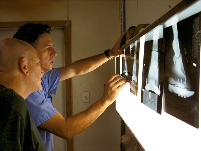
X-rays, ultrasound, MRIs and other diagnostic imaging methods might not often cross a farrier’s path. But when they do, they offer a tremendous opportunity to learn about the inner workings of the horse’s foot as well as information to help that particular case. At the fourth International Equine Conference on Laminitis and Diseases of the Foot, held Nov. 2-4 in West Palm Beach, Fla., several veterinarians offered discussions of various diagnostic imaging options for foot pain.
“The owner, farrier and veterinarian need solid information to treat the laminitic horse,” noted Bruce Lyle, a foot-focused veterinarian from Aubrey, Texas. This solid information is provided by specific radiology techniques, he said.
“The wishy-washy attitude of ‘some are going to get better regardless of what you do, and some are going to crater regardless of what you do’ (as an excuse for not taking radiographs) is counterproductive to the profession, the industry and most importantly the horse,” he warned.
Any diagnostic images must be taken with consistent, disciplined techniques, he noted. He described a radiographic protocol and schedule used by many clinicians who have experienced success in treating founder cases as well as other lamenesses, noting the following points: