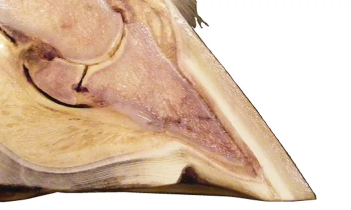
In my previous article (May/June 2014), I described how the perimeter of the sole has greater rigidity because of its attachment to the hoof wall, and provides a “ledge” for the solar border of P3 to rest on (Figure 1).
In some circumstances, deformation of the hoof wall and/or increased forces on P3 reduces the effectiveness of the ledge and the support it provides is either reduced or lost (Figure 2).

In this follow-up, I discuss this further with images to illustrate points.

Damage To The Ledge

Distortion or deviation of the distal wall can cause the border of P3 to rest on more flexible sole axial to (inside) the ledge. Figures 3a and 3b are radiographs of the left and right forefeet of a Quarter Horse that show this.

The loss of support of the ledge is most commonly and obviously seen in the chronic laminitis case, where separation of the laminae, disruption of the white line, dorsal wall deviation and rotation of P3 leave the bone’s tip, pressing directly on the sole (Figure 4). How successful or effective we are at treating acute laminitis cases will have a significant bearing on whether acute cases progress to the chronic state.

With low-grade laminitis caused by insulin resistance (IR), there is lengthening of the laminae and can be disruption of the white line, which weaken the support. The effect of these changes is made worse if the IR horse is overweight (Figure 5). Similar changes have been found in the feet of Brumbies (Australia’s feral horses) that travel great distances on hard substrate, causing low-grade mechanical laminitis.

SL_Fig_11

Figure 1

SL_Fig_11

Figure 2

SL_Fig_11

Figure 3a

SL_Fig_11

Figure 3b

SL_Fig_11

Figure 4

SL_Fig_11

Figure 5

SL_Fig_11

Figure 6

SL_Fig_11

Figure 7

The effectiveness of the ledge can be diminished due to a number of factors. We have limited control over some of these, such as genetic issues like weak walls and thin soles, and environmental factors such as wetness.

Figure 6 shows a Thoroughbred foot with a thin sole and flared wall, causing the sole to bend and P3 to “descend.” The inset shows the same foot further around the wall. This highlights the importance of considering the effectiveness of the ledge all around the foot.

There are other factors that we have a greater ability to control, such as so-called white line disease (Figure 7) and dietary factors that can affect hoof and white line integrity, such as a calcium-phosphorus ratio or zinc-copper ratio.

Aiding The Ledge

Trimming a “normal hoof” (with everything in the correct position) so the peripheral sole is weight-bearing helps maintain the effectiveness of the ledge. Figures 8a and 8b demonstrate this using barefoot and shod specimens.

SL_Fig_8a

Figure 8a

SL_Fig__8b

Figure 8b

SL_Fig_9

Figure 9

SL_Fig_10a_Reid

Figure 10a

SL_Fig_10b

Figure 10b

SL_Fig_11a

Figure 11a

SL_Fig_11b

Figure 11b

SL_Fig_12

Figure 12

SL_Fig_13_Maui

Figure 13

Regularly scheduled trimming also helps maintain the effectiveness of the ledge by limiting hoof distortion. However, even the over-grown foot can maintain ledge support if there is weight-sharing of the sole. The foot of a Brumby living on soft ground demonstrates this (Figure 9).

This weight-sharing of the sole by dirt (Figure 10a) can be replicated in different circumstances with the use of pads in boots, dental impression material under a pad, the use of Sole Guard, Styrofoam pads or EVA shoes. Figure 10b shows impression material on the sole after the shoe and pad are removed.

The force on the tip of P3 is greater in the upright foot (Figure 11a). Trimming the heels will reduce the bone angle and redistribute the horse’s weight more to the palmar foot and away from the toe (Figure 11b).

In some cases it is not possible to reduce the forces at the toe.

As the “foal hoof” grows out at the heel, the forces on the toe cause it to deviate (Figure 12). Complete removal of hoof wall distortion will leave the sole/ledge unsupported. The response to the pain caused by this can lead to contracture of the deep digital flexor muscle, often referred to as ballerina syndrome.

“Club feet,” which can’t have the heel trimmed down, are very often narrow feet. This allows for the hoof wall at the toe to be taken back to “ease breakover,” while leaving the ledge at the toe quarters to support P3. In these cases, where heel trimming may cause discomfort, greater trimming of the bars can potentially allow greater flexibility of the heels (Figure 13).

SL_Fig_14

Figure 14

SL_Fig__8b

Figure 15a

SL_Fig_9

Figure 15b

SL_Fig_10a_Reid

Figure 16

SL_Fig_10b

Figure 17

SL_Fig_11a

Figure 18

SL_Fig_11b

Figure 19

SL_Fig_12

Figure 20

In the true acute laminitis case, the white line formed before laminitis is triggered is, and remains, intact, so the ledge can continue to support P3 until it moves away due to hoof growth or hoof wall deviation. Figure 14 shows the intact white line in the foot of a Welsh Cob 6 weeks after severe acute laminitis. In this case the ledge continued to provide support even though P3 “sunk” in this foot within the first week.

In most situations, loss of P3 support from the ledge tends to occur over weeks and months, but over days and weeks following acute laminitis. Whatever trigger causes laminar detachment, the separation of the laminae appears to be due to the mechanical forces on P3 and the hoof wall. For this reason, maintaining the support of P3 by the ledge is extremely important.

For a case of acute laminitis, most hoof-care practitioners advise that the heels should be lowered, redistributing more of the horse’s weight to the palmar foot and away from the toe, and the toe trimmed to allow easier rollover at the toe, reducing the forces of separation on the laminae.

However, there is a wide variety of suggestions on how the feet should be managed following this trim, including barefoot, booted or shod. If shod, you must consider whether the case requires pads, sole support or frog support, or can be maintained with a low hoof/bone angle or with the heels wedged up.

I find an EVA shoe, or a wood or plastic Steward clog with solar impression material (Figure 15a), attached to the foot with a cast fulfills the requirements for dealing with acute laminitis cases. This solution provides sole support and reduces the forces of breakover around the foot, while the cast provides stability to the hoof capsule.

This method also has the advantage that the cast can be applied while the horse is standing on the clog (Figure 15b), thus minimizing the forces on the other foot, which can occur when other forms of support are applied. The EVA and cast can be seen on the radiograph of the foot of a horse with chronic laminitis.

Unfortunately, many cases of acute laminitis are seen in horses that have had low-grade laminitis due to IR or pituitary pars intermedia dysfunction that then progresses to clinical laminitis. These horses already have foot changes, with lengthened laminae and disrupted white line, so the effect of the ledge is already compromised and there is some distal descent of P3 within the hoof capsule.

The same principles of foot management apply to these cases, although, greater consideration must be given to solar support, to avoid pressure on the sole beneath the perimeter of P3, particularly at the toe (Figure 16).

There are now practical adaptations to the EVA shoe, including the addition of a leather pad with a leather rim (Figure 17), which can help with these concerns.

Trimming Practices To Avoid

Some trimming practices can reduce the support of the ledge. Removing any prominent rim of sole at the toe is likely to thin the sole and reduce the strength of the ledge, so should not be touched. The foot in Figure 18 has evidence of the effects of IR, with a widened “white line” (red arrow). After the sole was trimmed to this extent (blue arrow), this horse became lame and was very sensitive to pressure on the thinned sole.

Over-trimming of the hoof should be avoided (Figure 19). Removal of the distal wall so that it is no longer weight-bearing, takes away the part of the hoof that gives strength to the ledge and makes the sole weight-bearing instead of weight-sharing.

Traditionally, a heart-bar shoe has been used following hoof wall resections when dealing with laminitis or “white line disease” (Figure 20). Because the ledge is likely to already be compromised in these situations, consideration should be given to providing additional support to the sole and providing easier breakover to reduce the forces on the ledge.