“For something so small, equivalent in size to our little finger, the navicular bone can render a 1,000 pound, finely-tuned equine athlete into a pasture pet — permanently,” explains Dallas O. Goble, DVM, DACVS, former Director of Equine Clinics at the University of Tennessee and current head veterinarian for the Budweiser Clydesdale National Herd Health Program.

“As professionals in the field, you no doubt know, having had the misfortune to treat such a horse on occasion,” says the Strawberry Plains, Tenn., equine veterinarian. It seems that more clients’ horses experience navicular disease than ever before. Goble believes that farriers must understand the basic points of this crippling disease. By following his advice, you can give your clients a working knowledge of how to prevent, or at least manage, the damage before it’s too late.

The Navicular Bone Defined

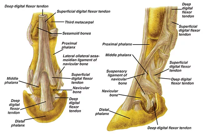
Of major importance to mobility of the limbs, Goble says the navicular bone “lays the groundwork of movement by serving as a conduit between the coffin bone and the deep digital flexor tendon (DDFT).” Defining it as a sesamoid bone (similar in nature to the sesamoid bones located behind the fetlock), it is positioned behind the coffin bone and below the small pastern bone, with the DDFT running underneath.

“As the navicular bone articulates with the coffin joint, or distal interphalangeal joint, on its anterior (front) surface to provide a smooth caudal (posterior) surface over which the deep flexor tendon glides,” says Goble. He further illustrates its impact by pointing to the tremendous forces placed on the bone when a horse gallops. “At a certain point in the gait, the horse essentially supports its entire weight with one leg.” During the initial phases of the stride, the loading is at a minimum as the limb flexes. However, as the stride progresses downward, loading takes place corresponding to the weight bearing on that limb.

Causes Of Navicular Disease

There is no single cause of navicular disease. However, Goble points to two significant contributors to this affliction: compression and tension.

The bone is supported by several ligaments above, below and on the side. This includes the impar ligament, which attaches the navicular bone to the coffin bone. With cartilage lying in between, as well as resting between the navicular bone and the DDFT, Goble contends that it is through repeated compression that the cartilage loses its shock absorbency, which he identifies as “the precursor to degeneration.”

Commonly seen along the flexor surface, continued cartilage erosion may eventually compromise the bone to the point that it becomes exposed. “Without this protective shield, the navicular bursa (a small sac that lies between the navicular bone and the DDFT), which is intended to prevent abrasion, also can become inflamed or damaged as a result of the constant friction.”

Excess tension placed on the supporting ligaments, the other major factor in navicular disease, is considered by many to be at the heart of the degeneration process. “Excess tension leads to strain on the impar ligament, inhibiting blood flow to the area. This leads to inflammation, resulting in a deficient supply of blood to the navicular bone,” explains Goble. He further maintains that if impar ligament strain becomes chronic, it can thicken and eventually reduce blood flow on a permanent basis.

“There are additional factors that can predispose a horse to injury, as well,” he continues. “Genetics contribute to bone density, weight of the horse in relation to foot size, conformation, hoof care, ground surfaces, weight of the rider and tack, the age of the horse and the list goes on.” Goble cites certain conformation imperfections, such as horses with short, straight pasterns, small feet, a downhill build and long toes with low heels as being particularly susceptible to navicular disease.

“The way the upright hoof is structured, for instance, limits the function of the frog and heel to absorb concession, thereby putting additional pressure on the heel region where the navicular bone lies. On the other hand, the long-toed-low-heeled horse is constantly stressing the navicular bone, regardless of whether he is standing or in motion,” Goble says, noting that this type of hoof conformation tends to have contracted heels, adding to the risk of damaging compression and strain.

The horse’s work can also play a role in Navicular Disease. Constant hill climbing, galloping and jumping puts additional strain on the DDFT, as well as causing overextension of the pastern and coffin joints. When coupled with traveling over hard or irregular surfaces that increase hoof concussion, the injury risk becomes much greater. “It is also possible that predominantly stabled horses, such as race horses or show horses, are equally as prone to navicular,” Goble noted. “Horses aren’t meant to stand still for long periods of a time. As a result, blood flow to the hoof slows while constant pressure on the navicular bones increase compression, a prescription for trouble.”

Symptoms

The initial symptoms are heel pain and mild and/or irregular lameness. The downward spiral begins with stress and inflammation of the ligaments that support the navicular bone. This is followed by reduced blood flow and hoof pressure and damage to the navicular bursa and/or DDFT with subsequent cartilage degeneration. Stumbling becomes frequent and the horse will appear to be on tiptoe or “walking on egg shells” as it tries to avoid putting pressure on the heels. “Lameness typically occurs in both front feet, although it is common for one foot to be worse than the other,” Goble states.

Reducing The Risks

“Advising your clients against allowing their horses to become overweight is essential to reducing the chances of developing navicular disease,” stresses Goble. “I consider a body condition score of 5.5 to 6.5 to be ideal for most horses. Secondly, proper foot care is mandatory. I don’t need to remind farriers of this. Yet, there is a perception by some owners that all horses have to be shod if they are to be ridden. That is a misconception. In my opinion, the horse should be shod according to the athletic work it performs, the needs of the individual hoof/horse itself and the surface over which it will be required to work.”

At the same time, he acknowledges that there are horses that must be shod to even be comfortable in a pasture environment. “And, of course, it almost goes without saying to recommend setting up a regular trimming/shoeing schedule — not just when there’s a problem,” he stresses.

Likely, your clients chose their horses based on size or color, rather than hoof conformation and quality. Still, Goble says you can remind them of a proverb that still holds true: “No hoof, no horse.”

Treatment

Goble believes treatment must be individualized to each horse. “There is no ‘one size fits all’ given that there is no one cause. As it is a progressively degenerative disease with the most advanced state believed to be non-reversible, there is a wide range of options available. The one constant, however, is shaping the foot into neurological and biomechanical balance, since navicular horses typically have long toes, underrun heels and thin hoof walls.”

Goble insists that the client, veterinarian and farrier should work as a team. “First, the veterinarian must determine the extent to which degeneration has taken place with a diagnostic work-up,” he says. “Next, with the client and farrier, a treatment plan must be decided upon and then put into place.”

In some cases, Goble prescribes medication, but other horses require a revised athletic program or minor management changes. Whatever the situation, he warns that initial treatment likely will need to be fine-tuned along the way.

Finally, Goble believes that early intervention is the key to controlling the damage. “As a farrier, you will be at the forefront of the situation should changes occur. Therefore, it would be in everyone’s best interest — particularly that of the horse — if you were to urge the client to keep you appraised of how the horse is responding. There is no substitute for working together when managing navicular disease. Seldom can one alone solve the problem.”