LEARN MORE

  • You can read a 2017 paper by Dr. Redden on principles for successfully sketching the equine foot.
  • Watch interview videos we recorded with Dr. Redden.

Access this content by visiting
AmericanFarriers.com/0718

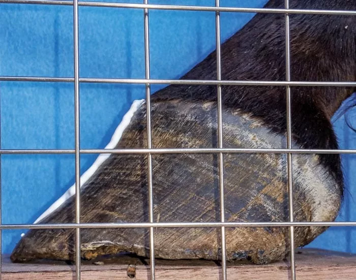
I am constantly striving to find ways to teach my veterinarian and farrier students how to tune their eyes to observe the smallest details. Many years ago, I learned that the caveman mentality is still a great way to teach. Simply studying the many messages left on stone by our predecessors from thousands of years ago allows us to step into their bare footprints and visualize what their eyes were seeing.

Also, one of the most effective methods of learning observation of the equine foot is through sketching. When we learn to draw what we see, we realize that our eye really did not capture the smaller details as the sketch may not even resemble what we thought we observed.

Farrier Takeaways

  • Using a grid can help you look at the foot in smaller sections, allowing for clearer recognition of subtle details.
  • When using the photo grid, move from one square to the next to help train your eye.
  • The practice of sketching the foot and using a photo grid can allow farriers and veterinarians to have more productive conversations with the footcare team through communicating on the same page.

Sketching The Foot

Our perspective of the foot varies greatly for several reasons. No one can see the world through another’s eyes. Therefore, the perspective views are different. We all are seeking quite different information because of our interests, reasons and goals. Our past experience demands more and more info as our learning curve goes through peaks and valleys.

I soon learned that the most informative perspective of the foot put our primary vision perpendicular to the subject of interest in the x,y,z planes. This view eliminates image distortion and offers consistent, repeatable, comparative views of the same foot at different times and sets a reliable standard for comparing the unique features of one to that of another.

This perspective view can produce images with photography, as well as radiographs. However, it is very difficult to study the foot looking at it in the non-distorted planes described. One would need to lie down on the ground, close one eye, position the other eye a couple inches off the ground and hope to see the image produced through the camera lens or the electrons striking the cassette. From my experience, that won’t happen. Therefore we must learn to produce consistent primary vision and beam orientation images to enhance our perspective view. Ultimately, this helps us learn to detect the smallest variances of distance. Here lies the key to better understanding of the subject.


The more we repeat sketching the unique characteristics of the toe, ground surface, heel bulb and pastern alignment, the more we detect …


Learning to capture what we see on paper opens the window of memory and allows us to reach a level where we no longer have to focus on every single aspect of the puzzle, as our eye and memory bank lets us zoom to the areas of highest interest following a micro-second scan of the image or radiograph. This learned quick scan is life’s learning curve that never ends. It continues to reach higher and higher levels of observation and interpretation. For finer points on sketching the foot, you can read my notes on the subject at Nanric.com/under stand_what_we_see.html.

The very reason for conflicting options many times is simply going to different schools. Visual perspectives are always different, as well as knowledge, experience and interpretive values. To help vets, farriers and owners communicate on the same page, we need common ground that brings our perspectives into a close resemblance so meaningful decisions can be made.

The growth ring patterns in Figures 1a and 1b allude to the 20-degree pastern angle. However, without the lateral radiograph, we would have no clue that half the coffin bone is gone. The grid allows us to simply count the 1-inch grid lines on the X-ray and transfer it to the photograph of the foot. Using this, plan the mechanics relative to the goals. In a case like this, pick up the rasp and leave the nippers in the box.

Examine the radiographs in Figures 2a and 2b. The location of the apex of the Lucent areas shows air density due to the multiple tracks from chronic bone disease and recurrent abscesses.

Smaller Portions

Breaking the foot down to smaller portions will benefit the view of the foot. We need to produce consistent, comparable photos and radiographic images. First, capture the foot using your method for consistency.

The next step uses a tool I created — a grid to superimpose over the photo or radiograph. Using the same perspectives, I then capture the same view using that superimposed grid. This tool allows the observer to break the image down into smaller areas.

My starter grid models have a 1-inch by 1-inch grid made with stainless steel welding rods that are aligned within a wood frame. Once the pen can follow the eye from one square to the next, the memory channels begin to record the relationship of the lines relative to other areas of the foot and once the outline or silhouettes complete, a new image is programmed.

With this foot in Figures 3a and 3b, the dish is the result of increased tension on the deep digital flexor tendon that is opposed by the excessive toe length. When looking at the image on the left, note the growth pattern, the pastern pushed forward and the heel bulb characteristics. The grid offers an easy way to break the outline (negative space around the foot) down into smaller focused areas.

The more experience that we gain by sketching the unique characteristics of the toe, ground surface, heel bulb and pastern alignment, the more we detect from what we missed prior. The better we draw, the more we see and the better our memory channels function. Once we reach a higher level of observation from the photos and radiographs, it will become instinctive to glance at a foot and visualize the radiographic image with reasonable certainty and confidence, and vice versa.

When we can feel the presence of the horse and visualize the cassette in place as the image was produced, we can then view it in that perpendicular relationship that we have been studying the photos in. What we see not only suddenly starts to make more sense, but what is missing from this picture also becomes more obvious.

Note that when left feet face west and right feet face east, it is quite easy to visualize the radiographic info as it relates to specific mechanical decisions. The flipped image that is programmed on many digital X-ray units automatically requires a mental flip that has no place in precision interpretation as it relates to specific trimming and/or shoeing goals. Viewing all images in the same plane they were made in is helpful.

Using the grid as an eye-training tool can greatly help the farrier detect the radiographic information and valuable external landmarks that he or she can use when shoeing a problematic foot on a sound athlete or an unsound foot on a horse with career- or life-threatening issues.

The grid can help veterinarians better understand the radiographic parameters that are constantly being altered by age, wear and tear, injury, disease and, last but not least, the alterations that occur due to routine or pathological shoeing.

The internal forces regulate the health of the outside. Farriers have the opportunity to learn how to appreciate and interpret the radiographic parameters that are altered with the swipe of the rasp, nippers and or shoe application. They also can greatly enhance their communication skills with the vets, trainers and owners, as well as increase their ability and more efficiently manage solutions.

This radiograph shows a cadaver foot that has been frozen for some time. Of interest is the Lucent zone along the dorsal face of the bone. This is the laminae zone. Note this makes up about half of the space between the bone and the outer wall.

This radiograph of a frozen cadaver foot reveals a uniform horn-lamellar zone down to the second wire. The hoof wall then has a distinct dish and healthy sole depth to the foot. This is slightly more sole than cup with a 5-degree pastern angle. These photographs and radiographs illustrate the overall value that helps farriers and veterinarians become more efficient in transferring the radiographic profile to the external landmarks. It is what we can’t see that is so very important.


July/August 2018 Issue Contents