Even those who have never had a CT scan are likely familiar with the process, which often entails a slow journey through a narrow tube. Given that the experience can elicit feelings of claustrophobia in human adults, it’s easy to imagine how complicated it can be to perform the same procedure on a 1,400-pound horse.
But what if the tube was no longer necessary?
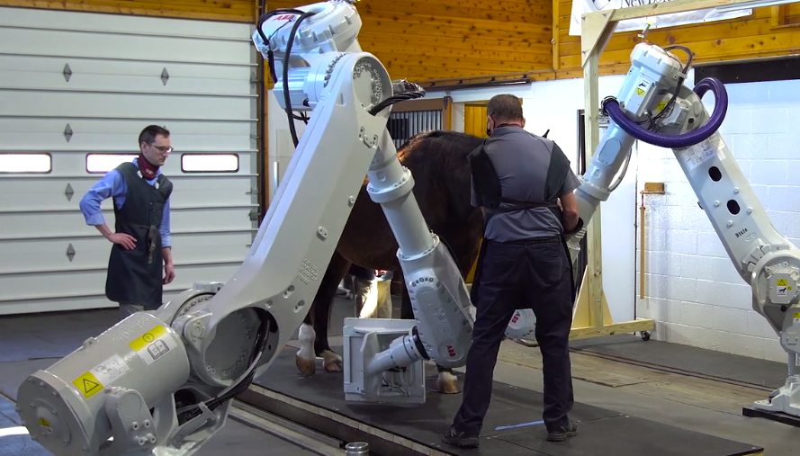
A partnership between the University of Pennsylvania School of Veterinary Medicine and the imaging technology company 4DDI is paving a new way to take medical scans of horses. Penn Vet is working with 4DDI to validate a new imaging system called Equimagine, which relies on robots that can move around a patient to collect high-resolution medical images.
Unlike traditional CT scanners, which require patients to be prone, the system can perform imaging studies on a standing patient. This means that a horse would not need to be anesthetized during the image collection.
“The reason this is so revolutionary is that the robots can easily move around the horse in any orientation,” says Barbara Dallap Schaer, medical director at Penn Vet’s New Bolton Center. “We can do the imaging in a patient that is standing and awake. From a clinical standpoint, we will see elements of the horse’s anatomy that we’ve never seen before.”
4DDI is a human imaging technology company, but feedback from veterinarians helped them see the potential value of an open-architecture imaging system for animal patients. Penn Vet is the first veterinary school to validate the system for clinical use, and will be working closely with the company to develop the relevant protocols.
“This collaboration with Penn is a great opportunity for us to improve our applications and work with the clinicians who will take them to the next level,” says Yiorgos Papaioannou, chief executive officer of 4DDI.
The robot-powered imager can collect not only typical, 2-dimensional CT images, but also fluoroscopic, or moving images; 3-dimensional images via tomosynthesis; and high-speed radiographs, capturing up to 16,000 frames per second. Eventually, researchers, clinicians, and engineers hope to be able to program the robots to capture images of a horse running on a treadmill, correcting for motion.
“The end results should be images that look very similar to traditional CT scan images without the risks, expense, and extra time required for general anesthesia and more diagnostic utility than conventional radiographs,” says Chris Ryan, radiologist at Penn Vet.
Dean Richardson, chief of large animal surgery at Penn Vet’s New Bolton Center, is looking forward to using the system to visualize portions of horse anatomy that cannot be seen with any other current imaging technique.
“One of the most important diseases of Thoroughbred racehorses is that they develop certain types of stress fractures that are very difficult to diagnose and characterize,” he says. “This technology has the potential to help diagnose those early enough that we can manage them and help prevent the horse from suffering a catastrophic breakdown on the race track.”
Beyond orthopedic applications, New Bolton Center researchers, clinicians, and even students will be involved in exploring the possible applications of the technology in a variety of specialities, including neurology, internal medicine, and sports medicine.
Such progress on the equine front will support significant applications in human medicine, notably for pediatric patients. Children who need CT scans often must be heavily sedated or in some cases put under general anesthesia in order to take images as they are too young to sit still for a test. If the machine itself can compensate for their movement, an image could be taken without the risk and complication of anesthesia.
“Instead of a child having to be anesthetized, they could sit there on their iPad and talk to their parents and have the image prepared in 30 seconds,” says Dallap Schaer. “That’s one of the translational pieces we hope to bring to Penn.”


