American Farriers Journal
American Farriers Journal is the “hands-on” magazine for professional farriers, equine veterinarians and horse care product and service buyers.

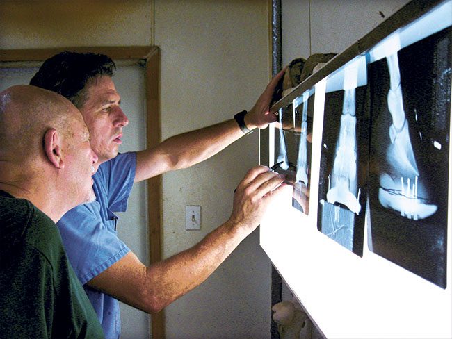
Dr. Bruce Lyle of Aubrey Equine Clinic in Aubrey, Texas, marks up an X-ray in this 2008 photo. Radiographs remain the most commonly used imaging modality in hoof care.
Many new diagnostic techniques and imaging modalities have come into use and it can often be challenging to understand which ones might be best suited for a given lameness problem. It is also important to know when to use these tools.
Unless the cause of lameness is obvious, a farrier should work with a veterinarian to determine which structures of the foot are involved. According to Dr. Jake Hersman of Animal Imaging, Irving, Texas, the most important part of any lameness work-up is still a basic clinical exam.
“The farrier’s initial look at the horse would evaluate the basic parameters of the horse’s conformation, heel angle, sole depth, medial-lateral balance, foot flight, hoof tester soreness, etc.,” says Hersman. “If the presenting lameness is still puzzling, however, localizing the soreness is extremely important before embarking on a shoeing plan. This is where the veterinarian needs to become involved — to localize the lameness with a series of nerve blocks, to further pinpoint the source.”
As a team, the farrier and veterinarian can find the problem and then devise a treatment plan for shoeing the horse for best results. Once the farrier and veterinarian have concluded their initial exams and lameness has been localized to the foot, the best way to locate and define the problem usually is diagnostic…